What is Dermo?
Dermo is caused by the single-celled protozoan parasite Perkinsus marinus that infects the hemocytes (blood cells) of the oyster. While not harmful to humans, Dermo can cause a range of problems in oysters, from sub-lethal infections that affect growth and fecundity to massive mortality events. Dermo infection can take up to two years to manifest in an individual. Below is a picture of a healthy (left) and infected (right) oyster.

Transmission
Dermo is most often spread through the disintegration of dead infected oysters. Uninfected oysters ingest active parasites released from dead infected oysters and become infected themselves. Dermo can be spread over long distances through live parasites in the water column. Transmission may also occur by scavengers feeding on dead infected oysters.
Dermo infections are most prevalent in warm, high salinity waters. The parasite proliferates in waters warmer than 25°C and salinities greater than 10‰. In the colder months of the year, Dermo can become dormant and therefore may be undetectable in oysters that are actually infected with the parasite.
Disease Monitoring
The Paynter Lab monitors for Dermo according to the practices of the Virginia Institute of Marine Science’s (VIMS) Shellfish Pathology Laboratory. The VIMS Shellfish Pathology Lab is a World Organization for Animal Health Reference Laboratory for perkinsosis (which includes Dermo) and is, therefore, the standard in Dermo monitoring.
During the fall three year check-in sampling season, before temperatures are too cold, populations are sampled for dermo testing. A random sample of 30-35 oysters are shucked and samples of the gill cross-section and anal tissue are removed per oyster. Tissues are placed in tubes with a Ray’s Fluid thioglycollate media for 5-10 days.
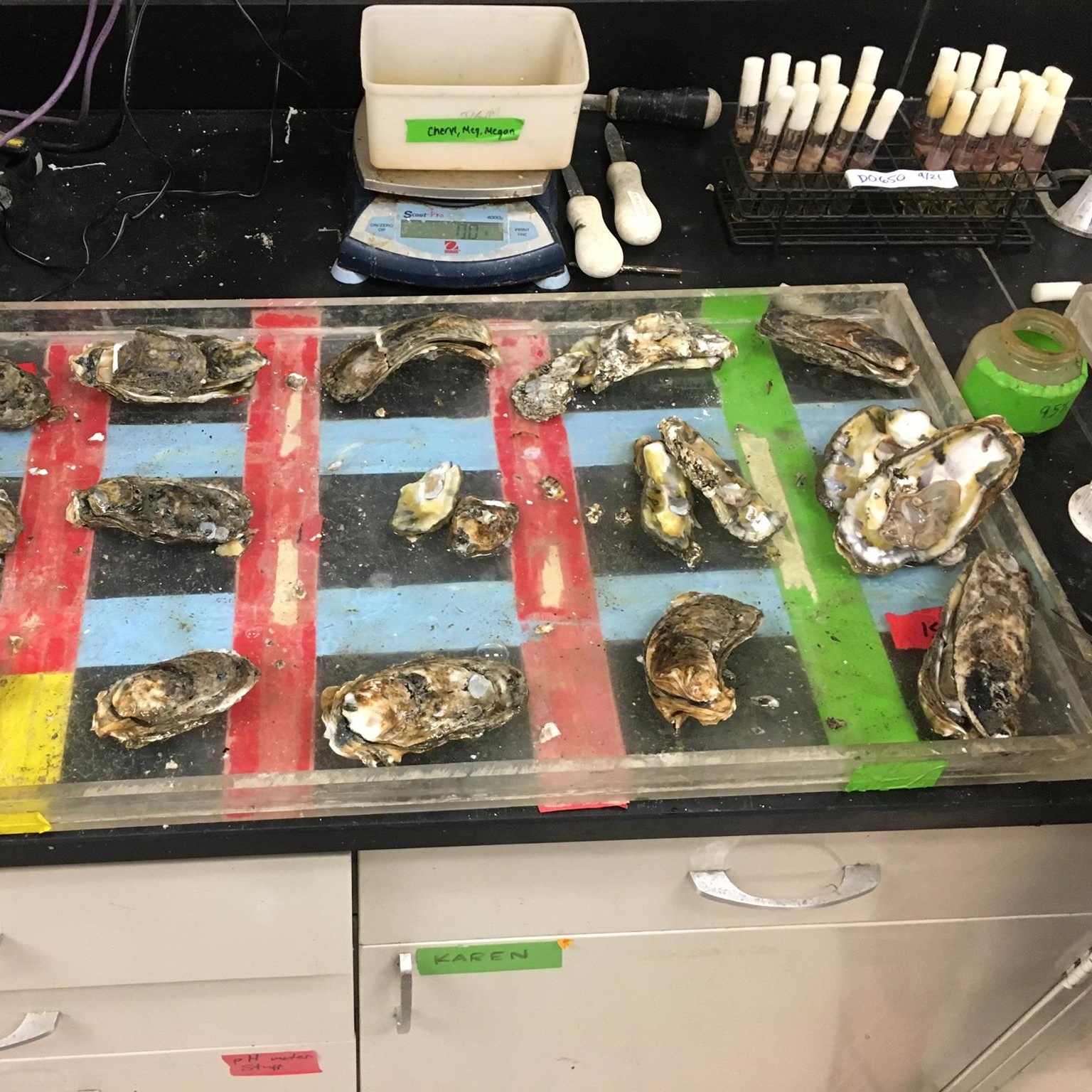

Oysters are separated out on platters where they are cleaned, weighed, measured, shucked, and dissected. Then the empty shell weight is collected to get the oyster meat weight.
After the incubation period, samples are examined under a microscope and Dermo cells are counted. Dermo infection levels are scored on a scale of 0 – 5, as indicated by the table below.


Microscopic images of oyster tissue after incubation period. Dermo cells are solid black spots in the tissue. Rare Dermo presence (left) vs Moderate Dermo presence (right).